Transcatheter ASD Closure in Thailand Your guide to cost, top specialists & hospitals
A hole between the heart's upper chambers can sit unnoticed for decades while the right side of the heart quietly takes the strain. It can often be closed through a vein in the leg.
What Is Transcatheter ASD Closure?
Also known as: Hole in the Heart Closure · Percutaneous Atrial Septal Defect (ASD) Device Closure
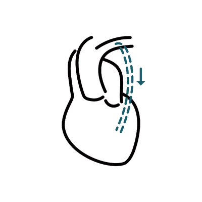
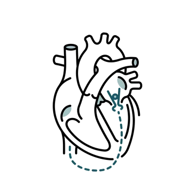
Transcatheter ASD closure is a catheter procedure that seals a hole between the heart's two upper chambers by delivering a small mesh device up through a vein in the leg. An atrial septal defect, or ASD, is a gap in the wall between the atria that has been there since birth.1 It lets blood cross from the left side of the heart to the right, so the right side and the lungs move more blood than they were built for. The procedure takes about an hour, guided by ultrasound, and leaves no chest incision.
Not every ASD needs closing. Small ones often cause nothing at all and are simply watched over the years. The defects that get treated tend to be the ones already stretching the right side of the heart, which an echocardiogram shows long before you feel much.
Closing the hole stops the extra flow, but it does not undo years of strain overnight. Breathlessness and fatigue usually ease over the months that follow, and how much of that recovers depends a good deal on how long the defect went unrepaired.
It can address a range of concerns, including:
Am I a Good Candidate for Transcatheter ASD Closure?
Suitability turns on where the hole sits, what the right side of the heart looks like, and what the pressure in the lung arteries is doing. None of those are things you can judge from symptoms.
A device needs somewhere to grip, so the anatomy decides the route long before anyone discusses a date.
Secundum type: Only defects in the middle of the wall are routinely suited to a device, which is roughly four in five cases.
An adequate rim: There has to be a firm border of tissue for the discs to clamp against on every side.
Measured properly: A transoesophageal or intracardiac ultrasound is what confirms this; a standard chest scan is not enough.
Closure is offered because of what the hole is doing to the heart, not because it is there.
Enlarged right chambers: A stretched right atrium and ventricle on echocardiography is the usual reason to intervene.
Symptoms that fit: Breathlessness on exertion, falling exercise tolerance, or palpitations that track back to the defect.
Small and silent defects: Many are simply monitored, and being watched rather than treated is a legitimate outcome.
Pressure in the lung circulation is the single finding that can turn closure from helpful into harmful.
Measured before deciding: Raised pulmonary pressures are assessed carefully, sometimes with a catheter study.
Severe elevation: Where pressure has risen far enough to reverse the direction of flow, closing the hole is not appropriate.
Borderline cases: These belong with an adult congenital heart team, not a general cardiology service.
A short list of checks keeps a low-risk procedure low risk.
No active infection: Any current infection is treated and cleared before a device is implanted.
Nickel sensitivity declared: Most devices contain a nickel titanium alloy, so a known allergy changes the choice.
Able to take aspirin: Six months of antiplatelet cover protects the device while your own tissue grows over it.
Who is not suitable for transcatheter asd closure?
- Severe pulmonary hypertension with flow reversed across the defect
- Primum, sinus venosus or coronary sinus defects, which need surgical repair
- A defect with a deficient rim that gives the device nothing to anchor against
- Active infection anywhere in the body until treated and cleared
- Unable to take antiplatelet medication for the six months after closure
Pricing
How Much Will Transcatheter ASD Closure Cost in Thailand?
What transcatheter asd closure costs at accredited hospitals in Thailand, and what your final quote depends on.
Get your exact price
Tell us what you're considering and we'll send a personalised quote from accredited clinics within 24 hours.
Get my free quoteTypical prices in Thailand
Typical ranges based on recent hospital quotes. Your exact price depends on the hospital and how complex your case is, and is confirmed at your teleconsultation.
Transcatheter ASD Closure
A small mesh device is threaded up to the heart through a vein in the leg to seal a hole between the two upper chambers, with no chest incision.
| Transcatheter ASD closure, fly-in package 2 nights Device closure of an atrial septal defect, catheter laboratory and two nights in hospital | $15,200 to $15,900 ฿517,500 to ฿540,500 |
Typically not included
- Consultation or treatment by a specialist outside the cardiac team
- Pre-operative medical clearance and pre-anaesthetic assessment
- Medication prescribed to take home after discharge
Get your exact price
Tell us what you're considering and we'll send a personalised quote from accredited clinics within 24 hours.
Hospitals Trusted for Transcatheter ASD Closure
From internationally accredited flagships to dedicated specialist hospitals, these are the kinds of facilities where international patients have this procedure.
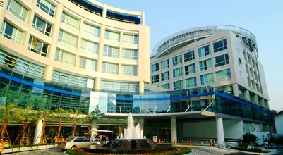
Bangkok Hospital
BDMS flagship tertiary campus with standalone heart, cancer, and neuro-orthopaedic hospitals.
View hospital profile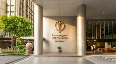
Bumrungrad International Hospital
Tertiary hospital with over 1,200 physicians treating 520,000+ international patients a year.
View hospital profile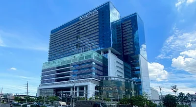
MedPark Hospital
Purpose-built tertiary hospital opened in 2020, focused on complex cardiac, cancer, and transplant care.
View hospital profile
Tell Us What You Need. We Do the Rest.
Share what you're considering and we'll come back with surgeon options, pricing, and a clear plan.
- Real hospital pricing with zero markup
- Matched with a specialist experienced in your specific procedure
- Full coordination from consultation to recovery


Trusted by patients worldwide
The complete guide to Transcatheter ASD Closure in Thailand
Everything below is for readers who want the full detail: costs broken down, types and techniques, recovery, risks and safety, and planning your trip.
Congenital and Structural Heart Teams in Thailand
A device closure is only as good as the team reading the septum. Structural heart work is its own subspecialty within interventional cardiology, and the distinction is worth checking.
What a Structural Heart Programme Looks Like
Device closure sits in the structural and congenital side of interventional cardiology, not general coronary work. A programme set up for it runs a catheter laboratory with three-dimensional transoesophageal echocardiography, an imaging cardiologist who scans alongside the operator, and cardiac surgical cover on site. Our partner hospitals in Bangkok run structural heart services of this kind rather than treating closures as an occasional case.
Experience That Matters Here
Ask about the number of septal closures the operator performs each year rather than catheter procedures in general, since the two are different skills. Fellowship training in structural or congenital intervention is the relevant credential. Adults with congenital heart disease are a distinct group with distinct problems, and a centre that sees them routinely will recognise the sinus venosus defect that a general service can miss.
Questions Worth Asking Before You Commit
Ask which device is proposed and why that one. Ask whether guidance will be transoesophageal or intracardiac, and what that means for your anaesthetic. Ask what the plan is if the rim turns out to be inadequate once you are on the table, because the honest answer is that the procedure is abandoned and surgery discussed, and a team that says so plainly is the team you want.
Understanding Your Results
The shunt stops immediately, but the heart takes months to unwind years of extra work. Knowing which changes are fast and which are slow makes the recovery far less unnerving.
What Changes Immediately
Blood stops crossing between the atria the moment the device is released, and the echocardiogram in the catheter laboratory shows it. Complete closure is achieved in the great majority of suitable secundum defects, with a small amount of residual flow in some cases that often disappears as tissue grows over the mesh. You will not feel this happen, which surprises people.
What Changes Over Months
The right side of the heart, stretched by years of extra volume, gets smaller again over the following six to twelve months. Breathlessness on exertion and exercise tolerance improve alongside that remodelling rather than immediately after the procedure. Results vary. Someone closed in their twenties tends to recover more completely than someone closed in their sixties, because there is less accumulated strain to reverse.
What Closure Does Not Fix
An arrhythmia that has already established itself, particularly atrial fibrillation, often continues after closure and needs treating in its own right. Raised pressure in the lung arteries may improve, may stabilise, or may not respond, depending on how far it has gone. Closure prevents further damage far more reliably than it reverses damage already done, and this is worth being clear-eyed about before you travel.
ASD Closure Cost in Thailand
What the Published Package Covers
Our partner hospital's cardiac centre quotes a package for transcatheter ASD closure of ฿517,500 to ฿540,500, valid to 31 December 2026 and covering two nights in hospital. The cardiac packages are more generous than the surgical ones in one specific way, which is that intermediate critical care and critical care unit time are named as included rather than charged if used. That matters on a procedure where the second night exists precisely to watch for rhythm problems.
What Sits Outside the Package
Pre-operative medical clearance and the pre-anaesthetic assessment are not in the price. Nor is a consultation with a specialist outside the cardiac team, which becomes relevant if another condition is picked up during the workup. Medication you take home after discharge is billed separately. Flights, accommodation and travel insurance are yours to arrange. Ask for the exclusions in writing before you travel, because a package price only helps you plan if you know its edges.
What Actually Moves the Total
The published range is narrow, which is unusual and reflects a procedure that varies less than most. The device is a single implant of a set design, the catheter laboratory time is fairly predictable, and the stay is fixed at two nights. The figures that change a quote are almost all found before the procedure rather than during it, in the imaging you need and in whether the pre-operative workup turns up anything that needs treating first.
How That Compares Internationally
Private ASD closure in the United States commonly runs from around $25,000 to $55,000, in Australia from about A$25,000 to A$50,000, and in the United Kingdom from roughly £15,000 to £30,000, though in the UK and Australia most closures happen inside the public system rather than privately. This is one hospital's rate rather than a Thai national price, and figures at other Thai hospitals differ in both directions.
Device Closure or Surgical Repair
Open surgical repair is the older route and remains the right one for a good number of people. The chest is opened, the heart is stopped on a bypass machine, and the hole is stitched closed or patched with a piece of the patient's own pericardium. It can close any defect regardless of where it sits or how big it is, it deals with a leaking valve or a misrouted pulmonary vein at the same time, and it leaves nothing implanted. The cost is a sternotomy, a longer hospital stay, and a recovery measured in weeks.
Device closure trades that reach for a much gentler recovery. There is no incision, no bypass machine, and most people are home in two days rather than a week. The limitation is real though. A device needs a rim of tissue to clamp against, so primum, sinus venosus and coronary sinus defects are not candidates, and very large secundum defects with a deficient rim often are not either. The decision is made on the transoesophageal scan, not on preference.
If your anatomy suits both, the catheter route is generally preferred for the simple reason that it asks less of you. If it does not, surgery is not a downgrade. It is the operation that fits the defect you actually have, and a congenital heart team that closes both ways is better placed to tell you which you are.
Types of Atrial Septal Defect
Where the hole sits in the wall decides more than its size does. Only one of the four types can usually be closed through a catheter, so the echocardiogram comes before any talk of a device.
Secundum ASD
A defect in the middle of the atrial wall, in the part known as the fossa ovalis. This is by far the most common type and the only one routinely suited to device closure, because there is usually a firm rim of tissue all the way round for the device to grip.
- Accounts for roughly four in five atrial septal defects1
- Sits centrally, so a double-disc device can clamp either side of the rim
- Closed through a leg vein under ultrasound guidance, with no chest incision
- Best for: centrally placed defects with an adequate rim on all sides
Primum ASD
A defect low in the atrial wall, near where the two valves between the upper and lower chambers meet. It frequently comes with a leaky mitral valve, so the valve and the hole are usually dealt with together in open surgery rather than through a catheter.
- Sits too low for a device to find a rim to anchor against
- Often paired with a cleft in the mitral valve that also needs repair
- Treated with open surgical repair rather than transcatheter closure
- Why it matters: a primum defect changes the operation, not just the approach
Sinus Venosus ASD
A defect high in the atrial wall, where the large vein returning blood from the upper body joins the heart. It is commonly linked to a pulmonary vein draining into the wrong chamber, which has to be rerouted at the same time.
- Frequently missed on a standard chest echocardiogram
- Usually accompanied by an abnormally connected pulmonary vein
- Repaired surgically so the vein can be redirected as well
- Why it matters: it is the type most often found late, on a transoesophageal scan
Coronary Sinus Defect
A rare defect involving the wall of the coronary sinus, the vein that drains the heart muscle itself. It allows a communication between the atria through that channel and is managed surgically.
- The least common of the four types
- Involves the drainage of the heart's own veins rather than the septum alone
- Surgical repair is the established route
- Why it matters: rarity makes an experienced congenital heart team more important, not less
How a Device Closure Is Done
The whole procedure happens inside the heart through a tube the width of a pencil. Here is what each stage involves and why it takes as long as it does.
Transoesophageal or Intracardiac Echo Guidance
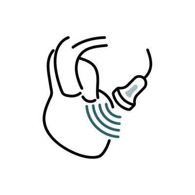
The cardiologist cannot see the defect on X-ray alone, so ultrasound does the seeing. Either a probe is passed down the gullet while you are asleep, giving a clear view of the wall from directly behind the heart, or a miniature ultrasound catheter is threaded into the heart itself. The scan measures the hole, checks the rim on every side, and confirms the device is sitting correctly before it is released.
- Measures the defect and its rim far more accurately than X-ray can
- Confirms the device position before the point of no return
- Intracardiac echo can avoid the need for a general anaesthetic
- Best for: every device closure, since none is done without it
Femoral Venous Access
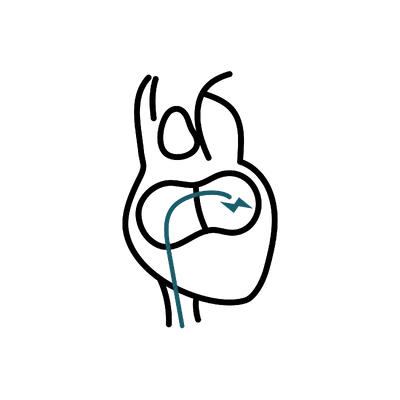
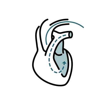
A short tube is placed in the femoral vein at the top of the leg and the equipment travels up the vein to the right atrium. Veins are lower pressure than arteries, so the access site is more forgiving than it is in a coronary procedure, and firm pressure afterwards is usually all that is needed.
- Uses a vein rather than an artery, which lowers the bleeding risk
- No incision beyond a puncture that closes with pressure
- Lying flat for a few hours afterwards protects the site
- Best for: the standard approach in almost all secundum closures
Balloon Sizing
A soft balloon is inflated across the hole to measure how wide it really is when stretched, since a defect is not a neat circle and its edges are flexible. The measurement decides the device size. Some operators size directly from the ultrasound instead, where the anatomy is straightforward.
- Establishes the stretched diameter, not just the resting one
- Guards against choosing a device that is too small to stay put
- Not always needed where echocardiographic measurements are clear
- Best for: defects with soft or irregular edges where sizing is less obvious
Double-Disc Device Deployment
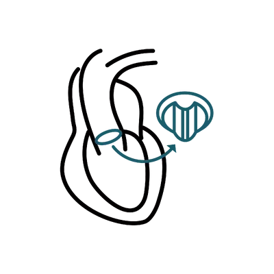
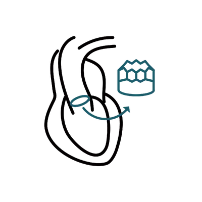
The device is a self-expanding mesh with a disc on each side and a waist between them. It is pushed through the tube in a collapsed state, one disc is opened on the far side of the wall, the other on the near side, and the two are drawn together so they clamp the septum. The cardiologist tugs it gently to check it is stable before releasing it.
- Clamps the wall rather than plugging the hole, which is what keeps it in place
- Can be recaptured and repositioned right up until it is released
- Heart tissue grows over the mesh in the months that follow
- Best for: secundum defects with a rim the discs can hold against
Recovery After ASD Device Closure
Day 1
You are monitored on the cardiac unit after the procedure, with heart rhythm and blood pressure watched continuously and the groin puncture checked for bleeding. You lie flat for a few hours so the vein seals, then sit up and walk short distances. An ECG and an echocardiogram confirm the device is where it was left.
Day 2
The second night in hospital is part of the package and exists to catch the small number of rhythm disturbances that show up early. A repeat echocardiogram checks the device position and looks for any residual flow across the wall. Aspirin is started, and most people are discharged with a written record of the device type and size.
Weeks 1–2
Walking and ordinary daily activity are encouraged from the start. Avoid heavy lifting and strenuous exercise while the groin site settles and the device begins to embed. A follow-up appointment reviews the access site and repeats the echocardiogram before you fly. Antibiotic cover is advised for dental work during this period.
Months 1–6
Your own heart tissue grows across the mesh over roughly six months, which is why aspirin usually continues for that long. Breathlessness and exercise tolerance improve gradually rather than suddenly as the right side of the heart remodels. A cardiologist at home takes over the follow-up echocardiograms, and most people return to full activity including competitive sport once cleared.
When Can You Fly After ASD Closure?
Most people are cleared to fly around seven to ten days after an uncomplicated closure, once the follow-up echocardiogram has confirmed the device is stable and the groin site has sealed. That is a little longer than the wait after a coronary stent, because the early days are when a rhythm disturbance or a device problem would show itself. Your cardiologist makes the call and can provide a letter for the airline.
When Can You Return to Work and Exercise?
Desk work usually resumes within a few days of discharge and walking is encouraged immediately. Heavy lifting and strenuous exercise wait about two weeks so the groin puncture is fully healed. Contact sport and competitive training are generally held for three to six months while your own tissue grows over the device, and are resumed on a cardiologist's clearance rather than on a fixed date.
Medication and Follow-Up Afterwards
Aspirin is standard for around six months, sometimes with a second antiplatelet for the first few weeks, to protect the device surface until it is covered by your own tissue. Antibiotic cover before dental treatment is advised for the first six months. Follow-up echocardiograms are usually done at intervals through the first year, and a cardiologist at home can carry these out with the written device record from Thailand.
Anaesthesia for ASD Closure
Most device closures are done under general anaesthesia, and the reason is the ultrasound rather than the catheter. Guidance usually comes from a transoesophageal probe passed down the gullet, which is uncomfortable to tolerate awake for the length of the procedure. You are asleep for about an hour to ninety minutes, and the puncture site in the groin is numbed with local anaesthetic as well.
Where the anatomy is straightforward, some centres guide the procedure with an intracardiac ultrasound catheter instead, threaded into the heart alongside the delivery system. That removes the need for the gullet probe, so the procedure can be done under sedation with you awake but relaxed. It is a decision the cardiologist and anaesthetist make together, based on the images they need and on your own airway and medical history.
A consultant anaesthetist stays with you throughout either way, monitoring heart rhythm, blood pressure and oxygen continuously. Waking is usually quick and most people are sitting up and eating within a few hours. A sore throat for a day or two is common after a gullet probe and settles on its own. Any tenderness afterwards is generally limited to the groin puncture and eases within a few days with simple pain relief.
Risks and Safety of Transcatheter ASD Closure
Device closure has a strong safety record in experienced congenital heart centres, and the serious complications are rare. They are worth understanding properly rather than glossing over, because a few of them declare themselves after you have gone home.
- Bruising or bleeding at the groin puncture, usually minor and settled with pressure
- Atrial arrhythmia in the days after closure, most often temporary
- Residual flow across the wall where closure is not complete
- Device embolisation, where the device shifts from position and has to be retrieved (rare)
- Erosion of the device through the heart wall, a rare but serious late complication
- Clot forming on the device surface before tissue has grown over it
- Allergic reaction in people sensitive to the nickel in the device alloy
- Air embolism or heart perforation during the procedure (uncommon in experienced hands)
Two things reduce your risk more than anything else. The first is honest sizing, which is why the transoesophageal scan matters as much as the catheter work. The second is taking the aspirin you are discharged with for the full period advised, because the device surface is only protected once your own tissue has grown across it.
Is ASD Closure Safe to Have in Thailand?
The safety of a device closure rests on the imaging and the operator rather than on the geography. What to establish before you commit is that the hospital has a dedicated congenital or structural heart programme, does transoesophageal-guided closures regularly, and has cardiac surgical cover available on site should a device need retrieving. Thailand's JCI-accredited cardiac centres use the same closure devices and imaging platforms available in Europe and North America.
How to Reduce Your Risk
Bring every scan you already have, since a defect measured twice by two teams is measured better. Make sure the plan includes a transoesophageal study rather than a chest echocardiogram alone, because rim assessment is what separates a device that holds from one that does not. Mention any nickel sensitivity before the device is chosen. Then take the aspirin for the full course you are given.
What About the Rare Late Complications?
Device erosion, where the device wears through the heart wall, is the complication that worries cardiologists most, and it is rare enough that most operators will never see one. It is more likely with an oversized device or a deficient rim, which is why sizing is done carefully rather than quickly. New chest pain or breathlessness in the weeks after closure is worth taking to an emergency department rather than waiting out.
Planning Your Trip to Thailand for ASD Closure
This trip is longer than a stent trip and shorter than an open-heart one. Most of the extra time is spent waiting to be cleared to fly rather than in hospital.
How Long to Stay in Thailand
Plan for 10 to 14 days. The first days cover your cardiology consultation, the transoesophageal echocardiogram that confirms the defect is closeable, and pre-procedure blood work. The procedure and two nights in hospital follow. The remaining week is follow-up, a repeat echocardiogram, and clearance to fly, which for most people lands around seven to ten days after the closure.
What to Bring With You
Bring every previous echocardiogram, ideally the images and not just the report, along with any cardiac MRI or CT, recent ECGs, and blood tests. If a defect has been measured before, that measurement is useful context even when it is repeated. Bring a list of your regular medications and note any nickel sensitivity, since it affects device choice rather than being a passing detail.
Travelling With Someone
Bring a companion if you can. You will have had a general anaesthetic, you will be told not to lift anything for a fortnight, and there are two nights in hospital where having someone who can speak to the team on your behalf is genuinely useful. Your care coordinator handles scheduling, hospital logistics and follow-up appointments across the stay.
Related Procedures
Other procedures that address similar goals or conditions, in case one of them is a closer fit for you.
Planning your treatment in Thailand
Independent guides to help you weigh the decision, before you commit to anything.
Common Questions About ASD Closure
Everything you need to know before your procedure

Medical References
Keep researching with AI
Open this guide in your AI assistant to dig deeper and ask the questions it didn't answer.
Opens in a new tab with this guide already loaded.
Medical disclaimer: Content on this site is provided for informational purposes and should not be treated as medical advice. Outcomes, timelines, and eligibility differ from person to person. Consult a qualified medical professional before making any decisions about surgery or treatment.
Ready to Get Started?
Speak with our care coordinators for a free, no-obligation consultation and personalised quote.
Speak to Our Team