Ptosis Correction in Thailand Your guide to cost, top specialists & hospitals
Repositioning the muscle that lifts your eyelid so it stays where it should and your full field of vision returns.
What Is Ptosis Correction?
Also known as: Droopy Eyelid Surgery · Surgical Repair of Eyelid Ptosis
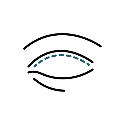
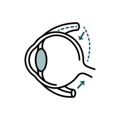
Ptosis correction is eyelid surgery that lifts a drooping upper lid by tightening or reattaching the levator, the muscle that raises the eyelid, or its stretched tendon. It sets the lid to a height that clears the pupil, widening your visual field and easing the constant brow-raising many people use to see past a low lid. The repair is usually done through a hidden crease incision, or from behind the lid in milder cases, and tends to last for years. Most operations take 45 to 90 minutes under local anaesthesia, so the surgeon can set the height while you open your eyes.
A droopy lid creeps in slowly and is easy to live with for too long. The cause matters as much as the droop, so your surgeon measures how far the muscle still travels before choosing how to lift it.
Matching the two lids closely is the aim, and for most people the change looks natural. Symmetry to the millimetre cannot be promised, which is why the height is set while you are awake and reviewed once swelling settles.
It can address a range of concerns, including:
Am I a Good Candidate for Ptosis Correction?
Suitability for ptosis repair comes down to one key measurement, a confirmed cause, and an eye surface that can tolerate a higher lid.
How far your lid muscle travels is the single number that determines the right technique.
5mm or more of excursion: Reasonable levator function points to levator advancement, the standard repair through the eyelid crease with the lid height set while you are awake.
Under 4mm of function: Very poor function, typical of severe congenital ptosis, needs a frontalis sling that bypasses the muscle entirely.
Mild droop, positive phenylephrine test: A 1-2mm droop that lifts with test drops may suit the internal MMCR approach, which leaves no visible scar.
Specialists confirm why the lid is drooping before planning how to lift it.
Aponeurotic or congenital ptosis confirmed: Age-related stretching of the levator tendon and congenital muscle weakness are the standard surgical cases.
Neuromuscular causes excluded: A droop that fluctuates through the day can signal myasthenia gravis, which needs medical workup before any surgical plan.
Thyroid eye disease stable: If lid position is still changing month to month, surgical measurements become unreliable and the operation waits.
Previous eyelid surgery flagged: A lid operated on before carries scarred levator tissue, so it is treated as a separate revision category where technique choice and the predictability of the result both differ.
Lifting the lid exposes more of the eye, so the surface has to be able to cope.
Adequate tear film: Significant dry eye raises exposure risk once the lid sits higher, so it is assessed and managed first.
A good Bell's reflex: The eye's protective upward roll during closure is checked, because a poor reflex compounds any exposure problem.
A quiet, healthy lid: No active lid infection and stable ocular health before surgery is scheduled.
Symmetry to the millimetre is the goal, but biology does not always cooperate fully.
Near-symmetry, not perfection: Most patients achieve a significant, natural-looking correction; sub-millimetre matching between two lids cannot be promised.
Awake surgery is the safeguard: Local anaesthesia lets the surgeon set lid height with your cooperation during the operation, the most reliable route to a balanced result.
Three months to judge: Early height differences usually settle as swelling resolves; the position at month three is essentially the final outcome.
Who is not suitable for ptosis correction?
- Levator function not yet formally measured
- Suspected myasthenia gravis or a fluctuating droop awaiting workup
- Thyroid eye disease still changing month to month
- Significant dry eye or a poor Bell's reflex not yet assessed
- Smokers unwilling to stop at least four weeks before surgery
Pricing
How Much Will Ptosis Correction Cost in Thailand?
What ptosis correction costs at accredited hospitals in Thailand, and what your final quote depends on.
Get your exact price
Tell us what you're considering and we'll send a personalised quote from accredited hospitals within 24 hours.
Get a Free QuoteTypical prices in Thailand
Typical ranges based on recent hospital quotes. Your exact price depends on the hospital and how complex your case is, and is confirmed at your teleconsultation.
Ptosis Correction
The drooping upper eyelid is lifted by tightening or reattaching the levator, the muscle that raises the lid, so it clears the pupil and widens your field of vision.
| Correction of ptosis by levator correction, two eyes Day case Local anaesthetic, outpatient, no overnight stay | $2,450 ฿83,000 |
Typically not included
- Correction of one eye only, which is not what this package covers and has to be quoted separately
- Frontalis sling surgery, which is a different operation for eyelids with poor levator function, and any fascia lata harvest it needs
- Revision of a previous ptosis repair
- Any hospital admission or overnight stay, since the rate is for an outpatient day case
- Levator function measurement and phenylephrine testing before surgery, along with laboratory tests and imaging
- Pre-operative medical clearance and fitness assessment
Get your exact price
Tell us what you're considering and we'll send a personalised quote from accredited hospitals within 24 hours.
Hospitals Trusted for Ptosis Correction
From internationally accredited flagships to dedicated specialist hospitals, these are the kinds of facilities where international patients have this procedure.
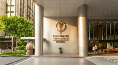
Bumrungrad International Hospital
Full-service hospital with over 1,200 physicians treating 520,000+ international patients a year.
View hospital profile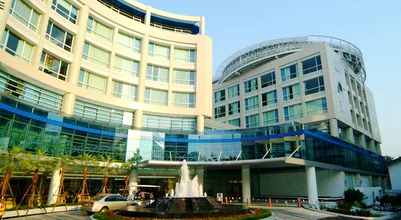
Bangkok Hospital
BDMS flagship hospital campus with standalone heart, cancer, and neuro-orthopaedic hospitals.
View hospital profile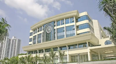
Samitivej Sukhumvit Hospital
Full-service hospital known for paediatrics, home to Thailand's first private children's hospital.
View hospital profile
Tell Us What You Need. We Do the Rest.
Share what you're considering and we'll come back with surgeon options, pricing, and a clear plan.
- Real hospital pricing with zero markup
- Matched with a specialist experienced in your specific procedure
- Full coordination from consultation to recovery


Trusted by patients worldwide
The complete guide to Ptosis Correction in Thailand
Everything below is for readers who want the full detail: costs broken down, types and techniques, recovery, risks and safety, and planning your trip.
Ptosis Surgeons & Clinics in Thailand
Ptosis repair demands oculoplastic subspecialist training and judgment. Here is what sets our partner centres apart.
Leading Oculoplastic Centres in Bangkok
Our partner hospitals have dedicated oculoplastic departments within their ophthalmology divisions. Equipment includes ophthalmic microsurgical instruments, high-resolution photography for lid height documentation, and facilities for both levator repair and frontalis sling under local or general anaesthesia.
Experienced Ptosis Surgeons
Our partner surgeons completed oculoplastic fellowships and perform ptosis repair (both adult and paediatric) as a regular part of their surgical practice. They manage all complexity levels, from straightforward aponeurotic ptosis to congenital cases requiring frontalis sling and bilateral adjustments.
What to Look for in a Ptosis Surgeon
Confirm they perform ptosis repair under local anaesthesia with intra-operative lid height adjustment; this is the most reliable approach for precision. Ask about their approach to bilateral cases and how they manage asymmetry. Check before-and-after photographs of cases similar to yours, paying particular attention to the symmetry achieved.
Understanding Your Results
Ptosis correction produces a measurable change in lid position and visual field. Here is what realistic results look like.
Typical Results
Successful ptosis repair restores the lid to a height that clears the pupil and matches the fellow eye. Patients report elimination of compensatory brow-raising, relief from forehead headaches, and a wider, more open visual field. The correction is durable; age-related recurrence can occur after many years, but a well-performed repair lasts a long time.1,3
What Results Can You Expect?
Perfect symmetry is the goal but not always achievable to the sub-millimetre level. Your surgeon will explain what degree of correction is likely based on your levator function and the severity of the droop. In the vast majority of cases, the improvement is significant and the result looks natural. Post-operative photographs and repeat visual field testing document the change objectively.
Ptosis Correction Cost in Thailand
Average Cost of Ptosis Correction
We hold one fixed fly-in package price for ptosis correction at our partner hospital, valid to 31 December 2026. It is ฿83,000, roughly $2,441, for correction by levator correction in two eyes, under local anaesthetic as an outpatient with no overnight stay. The wording matters more than the number. This is a two-eye price, so if only one lid has dropped you are not buying this package and the single-eye figure has to be quoted to you. This is the rate we hold at one hospital rather than a national Thai price, and figures across the same network vary noticeably by branch.
Cost Breakdown
The package covers the oculoplastic surgeon, the local anaesthetic, the operating theatre and the outpatient stay, for both eyelids. Outside it sit the assessment that decides whether you need this operation at all, meaning levator function measurement and phenylephrine testing, along with pre-operative medical clearance, laboratory tests and imaging, consultant fees from other departments, and medication for conditions you already take. A frontalis sling, which is the different operation used when the lifting muscle has too little power for levator correction, is not covered by this package and neither is a fascia lata harvest if autogenous tissue is used.
What Affects the Price?
The published figure does not flex, so what changes your total is whether the operation you need is the one being priced. Levator correction on both lids is the case this package fits. One lid only, a frontalis sling for poor levator function, or a revision after previous surgery elsewhere all sit outside it and are quoted individually. It is worth asking for the one-eye figure early even if you expect to need both, because it tells you how the hospital prices the operation rather than the package, and it is the number that makes a single-lid quote comparable with anything you have been given at home.
Thailand vs International Price Comparison
The package we hold is ฿83,000, about $2,441 for two eyelids, against $5,000 to $8,000 in the US, A$4,600 to A$7,600 in Australia and £4,000 to £7,000 in the UK. Check what the comparator covers before you weigh it, because Western quotes for ptosis are commonly written per eyelid, which would make the gap wider than the raw figures suggest. The surgical techniques and instruments used at our partner hospitals are identical to those at leading oculoplastic centres worldwide.
Surgical vs Non-Surgical Ptosis Treatment
A prescription eye drop containing oxymetazoline (sold as Upneeq) is the main non-surgical option for a mild droop. It works by stimulating Muller's muscle to contract, lifting the upper lid by a small amount.1 It can be a useful daily lift for someone with very slight ptosis, and the same response is the basis of the phenylephrine test your surgeon may use to predict how a Muller's muscle procedure would behave.
The limits are significant, though. The drop lifts only a fraction of what surgery achieves, lasts a single day, and must be re-dosed every morning indefinitely, so the cost and routine never stop. It does nothing for moderate or severe ptosis, nothing for poor levator function, and nothing for a congenital droop, and it cannot correct asymmetry with any precision. It treats the appearance for a few hours rather than the stretched or detached muscle causing the problem.
Where the lid genuinely covers the pupil, narrows your field of vision, or sits clearly lower than the other eye, surgery is the route that fixes the underlying muscle and holds for years rather than hours. Repositioning or reattaching the levator, with the height set while you are awake, is what delivers a lasting, matched result, and that is what the rest of this page covers.
Types of Ptosis Correction
The choice of technique depends almost entirely on levator function, how many millimetres of excursion the muscle produces from downgaze to upgaze. Good function means the muscle can be advanced or shortened directly. Poor function means it needs to be bypassed.
Levator Advancement or Resection
The standard approach for acquired ptosis where the levator still has reasonable function. Through an eyelid crease incision, the surgeon identifies the stretched aponeurosis and reattaches or shortens it. Lid height is set under local anaesthesia with patient cooperation.
- Most widely used technique for moderate to good levator function
- Intra-operative adjustment allows precise lid height matching between both eyes
- Incision hidden within the natural eyelid crease
- Best for: age-related or aponeurotic ptosis with at least 5 mm of levator excursion
Frontalis Sling
When the levator muscle has very poor function, typically in congenital ptosis, a sling connects the eyelid to the frontalis muscle of the forehead, allowing the brow to open the eye. The sling material can be autogenous fascia lata or synthetic material depending on the patient's age.
- Bypasses the non-functioning levator entirely
- Uses the forehead muscle to lift the eyelid via a suspensory connection
- Fascia lata harvest is preferred in older children and adults for long-term durability
- Best for: severe congenital ptosis with levator function under 4 mm
Ptosis Correction Techniques
Technique selection is driven by levator function measurements. Pre-operative testing records the distance your lid travels from downgaze to upgaze and the degree of droop from pupil centre to lid margin. These numbers determine which approach will produce the most reliable result.
Muller Muscle-Conjunctival Resection (MMCR)
An internal approach that accesses the lid from beneath, shortening the Muller muscle with a strip of conjunctiva. No external skin incision. Reserved for mild ptosis cases where phenylephrine drops confirm the Muller muscle alone can achieve the required lift.
- No external incision, no visible scar
- Highly predictable in carefully selected mild cases
- Shorter operating time with faster post-operative recovery
- Best for: mild ptosis (1–2 mm) with positive phenylephrine test
Adjustable Suture Technique
The levator is reattached using sutures that can be adjusted at the slit lamp in the hours after surgery. If the lid height is not quite right once swelling and anaesthetic effects settle, the surgeon repositions the suture without returning to theatre. Adds a precision safety net.
- Allows fine-tuning of lid height after the initial repair
- Adjustment performed at the slit lamp, no second trip to theatre
- Reduces the risk of under- or over-correction requiring formal revision
- Best for: complex or bilateral ptosis where achieving exact symmetry is critical
Fasanella-Servat Procedure
A long-established internal technique that removes a small strip of the upper tarsus along with conjunctiva and Muller muscle, shortening the lid lifting structures from behind. Like other posterior approaches it leaves no external scar, and it suits mild ptosis where the levator already works well. The amount removed is set against the millimetres of lift needed.
- Internal approach with no visible external skin incision
- Reliable, well-documented option for small, fixed amounts of lift
- Shorter procedure with a quick, comfortable recovery
- Best for: mild ptosis (1-2 mm) with good levator function
Ptosis Correction Recovery Timeline
Days 1–3
Swelling and bruising around the operated lid peak within the first 48 hours. The lid may initially sit higher than intended; this settles as swelling resolves. Cold compresses, head elevation, and prescribed lubricating drops keep you comfortable. Daily check-ins monitor early lid position. If a frontalis sling used autogenous fascia lata, you also have a small thigh wound that is sore to walk on for the first few days; keep it clean and dry as directed.
Days 5–7
Sutures are removed if an external approach was used. Bruising begins to fade and initial swelling decreases. Your surgeon assesses early lid height and symmetry and provides guidance on returning to light daily activities. A fascia lata thigh wound is also checked at this point; walking discomfort eases over the first one to two weeks.
Weeks 2–4
Visible bruising and swelling resolve. The lid settles toward its corrected position and feels progressively more natural. Desk work and normal daily routines can resume. Avoid straining and heavy lifting until given clearance.
Months 1–3
Lid height and contour reach their stable position as residual oedema fully clears. Visual field improvement is complete. The symmetry between your eyelids settles to its long-term result; what you see at three months is essentially the final outcome.
When Can You Fly After Ptosis Correction?
Most patients fly home 7–10 days after surgery, once sutures are removed and the surgeon confirms the lid is healing in correct position. Cabin pressure does not affect the repair. Use lubricating drops during the flight to protect the eye from dry cabin air.
When Can You Return to Work and Exercise?
Desk work can resume within a week once sutures are out. Light walking is encouraged from day one. Avoid gym workouts, heavy lifting, and anything that significantly raises blood pressure for 3–4 weeks. Contact sports should wait until your surgeon gives full clearance. If your frontalis sling used autogenous fascia lata, expect a sore thigh wound that makes walking uncomfortable for the first 1–2 weeks; stairs and longer walks come back gradually as the donor site heals.
When Will You See the Final Result?
The lid may sit slightly high initially due to swelling; this is normal and settles over 2–4 weeks. By three months, residual oedema has fully resolved and the lid height has stabilised to its permanent position. Most patients see their final symmetry between months two and three.
Anaesthesia for Ptosis Correction
Ptosis correction is done under local anaesthesia with light sedation, so you stay awake but relaxed and feel no pain.1,2 The surgeon numbs the eyelid with a small injection, and the sedation keeps you calm and comfortable throughout. Staying awake is deliberate rather than incidental: at a key point the surgeon asks you to open your eyes and look in different directions so the lid height can be set and matched to the other eye while you are sitting up. A fully asleep patient cannot do this, which is why awake surgery gives the most reliable symmetry.
You are looked after by the surgical team the whole time, with your comfort and observations monitored throughout the short procedure. You will not see the surgery happening: the eye being operated on is numb and shielded, and your view is of the room rather than the instruments. Most people are surprised by how undramatic it feels, more like lying still with a slight sense of pressure and tugging than anything sharp.
Before surgery you have an assessment that includes your levator function measurements and a review of any medications, particularly blood thinners, which need pausing beforehand. During the operation you genuinely feel nothing in the eyelid itself. Afterwards there is some mild soreness, tightness, and watering for a few days, which settles quickly and is well managed with cold compresses, lubricating drops, and the pain relief your surgeon prescribes.
Risks and Safety of Ptosis Correction
Ptosis repair is technically demanding because achieving exact symmetry between two eyelids is one of the most precise goals in oculoplastic surgery. Understanding the risks helps set appropriate expectations.
- Residual asymmetry between the two lid heights, the most discussed risk2
- Over-correction leaving the lid too high with visible sclera above the iris
- Under-correction requiring a secondary adjustment procedure
- Lagophthalmos, the lid not closing fully so the cornea is left exposed, which can cause dryness, grittiness, or exposure keratopathy and needs prompt attention1,2
- Temporary dry eye symptoms from altered lid dynamics, usually self-limiting
- Post-operative swelling and bruising, expected and resolves within weeks2
- Infection or haematoma, rare with proper technique
- Thigh donor-site problems (wound haematoma, scar, or temporary leg discomfort) in frontalis sling cases where autogenous fascia lata is harvested
Intra-operative lid height assessment under local anaesthesia, careful pre-operative levator function testing, and adjustable suture techniques all reduce the risk of asymmetry. A thorough examination also identifies factors like dry eye disease or poor Bell's reflex that could influence the surgical plan.
Is Ptosis Correction Safe in Thailand?
Yes. Our partner eye centres hold JCI accreditation and employ oculoplastic surgeons, ophthalmologists with fellowship training in eyelid and orbital surgery. Ptosis repair is a core procedure at these centres, not an occasional case. The safety profile is consistent with published outcomes from international oculoplastic units.
How to Reduce Risks
The most effective risk-reduction step is choosing an oculoplastic surgeon who performs ptosis repair regularly and uses intra-operative lid height adjustment under local anaesthesia. Pre-operative levator function testing ensures the correct technique is selected. Adjustable sutures provide an additional safety margin for fine-tuning in the hours after surgery.
When Is Revision Needed?
Minor asymmetry in the early weeks is common and usually resolves as swelling settles. Persistent asymmetry beyond three months may warrant a small revision procedure, typically a straightforward adjustment under local anaesthesia. Over-correction causing lid retraction is less common but may require surgical lowering. Choosing an experienced oculoplastic surgeon reduces the need for revision considerably.
Planning Your Trip to Thailand for Ptosis Correction
Ptosis correction requires 7–10 days in Thailand, a short trip for a procedure with long-lasting impact.
How Long to Stay in Thailand
Plan for 7–10 days. Your first day covers the oculoplastic assessment, levator function testing, and surgical planning. Surgery follows within 1–2 days. Suture removal at day 5–7 and a final lid height check complete the visit before you fly home.
What Is Included in a Medical Trip
Your care coordinator manages scheduling, hospital transfers, and all follow-up appointments. The surgical quote covers the oculoplastic surgeon, anaesthesia, operating theatre, nursing care, medications, lubricating drops, and suture removal. A detailed surgical summary is prepared for your home ophthalmologist.
Recovery in Bangkok
Stay near your hospital for the first week. Ptosis recovery is straightforward and you are mobile from day one, but the suture removal and lid height assessment need to happen on schedule. Your coordinator can suggest nearby hotels that are practical for medical recovery stays.
Related Procedures
Other procedures that address similar goals or conditions, in case one of them is a closer fit for you.
Planning your treatment in Thailand
Independent guides to help you weigh the decision, before you commit to anything.
Common Questions About Ptosis Correction in Thailand
What patients ask about ptosis repair

Medical References
Keep researching with AI
Open this guide in your AI assistant to dig deeper and ask the questions it didn't answer.
Opens in a new tab with this guide already loaded.
Medical disclaimer: Content on this site is provided for informational purposes and should not be treated as medical advice. Outcomes, timelines, and eligibility differ from person to person. Consult a qualified medical professional before making any decisions about surgery or treatment.
Ready to Get Started?
Speak with our care coordinators for a free consultation and find out if ptosis correction can restore your natural eyelid height and visual field.
Get a Free QuoteEyelid & Reconstructive